
As described in a recent post, dental conditions are often hidden and painful. The following posts are going to help describe conditions that pets can get as well as treatment. Many people I talk with are surprised how we can help pets with dental conditions and save teeth whenever possible.
While only 6% of malignant tumors of the body occur in the mouth, additional growths classified as benign occur. The trouble with the benign classification is that although they may not spread to other parts of the body, they are often locally invasive, painful, and destructive. The other trouble with both benign, benign-locally invasive, and malignant tumors is that many types of them look quite similar. As stated in the prior section, all types of gingival enlargements should be biopsied to help ensure the best treatment. Biopsy samples should be sent to an ORAL pathologist. Dr. Cindy Bell (KSU) currently is the leader oral pathology. Speaking from experience, it appears her reads tend to be more definitive than others. She is down to earth and will actually talk to veterinarians about treatment or help provide connections to speak to a veterinary dentist about the case.
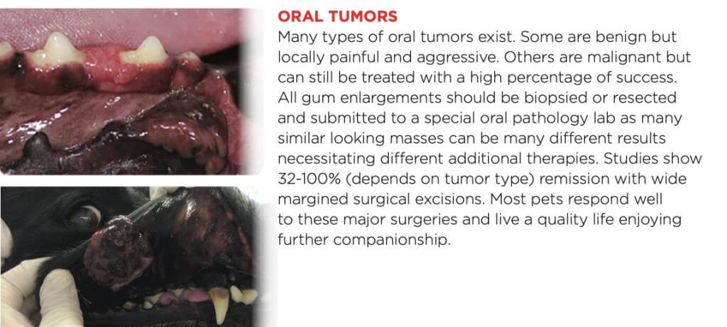
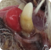
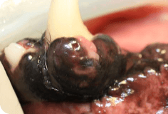
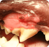
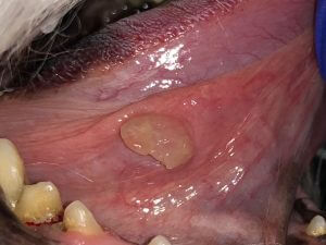
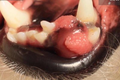
Classifications of the following image growths may be plasma cell tumor, peripheral odontogenic fibroma, focal fibrous hyperplasia, papillary squamous cell carcinoma, lymphoma, pyogenic granuloma, canine acanthomatous ameloblastoma. I’m sure there are a few I’ve missed as well. Take note – the word epulis was not used to describe any of the growths. Epulis is a poor descriptive term. Most previously classified epulides are either peripheral odontogenic fibromas or ossifying fibromas. Also note, that all of the below images look similar and most of them are severe, locally invasive, or symptoms of additional disease.
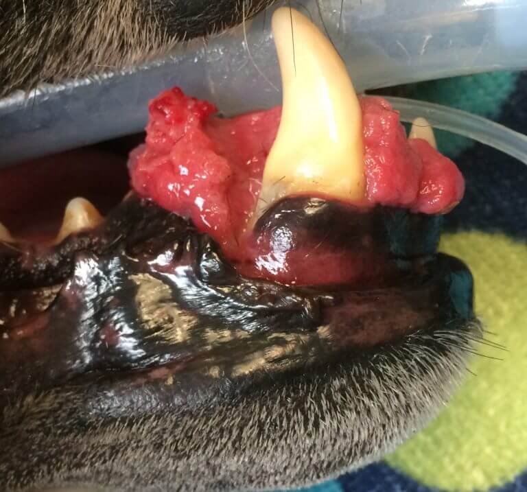
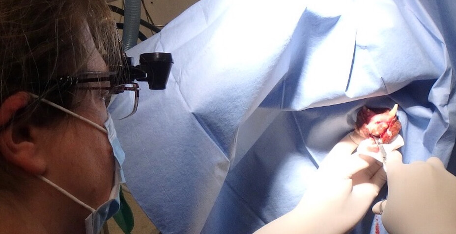
Many of these tumors require wide margined surgical excision; meaning large parts of the jaw(s) are removed. When taking off parts of the upper jaw, large holes can be made but the lip can cover and make the outcome be quite cosmetic for many cases.
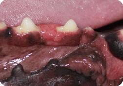
A mandibulectomy finish is shown below.
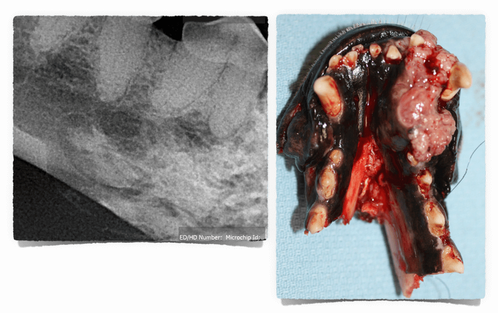
Studies show client satisfaction following mandibulectomies and maxillectomies in dogs is 85%, and was proportional to the postoperative survival time. It has been thought that cats do not do well after these types of surgeries. Surprising to many, a similar percentage of clients with cats after a mandibulectomy procedure said they would choose the same course of action given the circumstance despite the fact that 75% of the cats had mild to moderate adverse effects (tongue location, drooling, difficulty grooming, jaw alignment issues) for the rest of their lives.
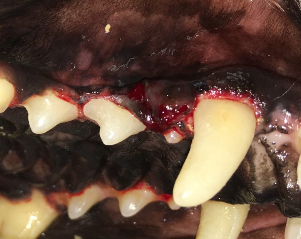
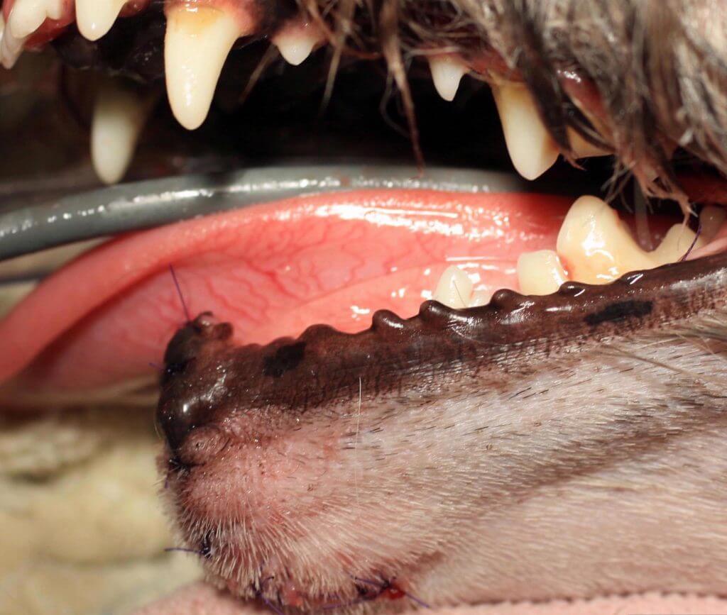
This mass (behind the canine and is subtle) was found very early and had a great outcome. Other larger ones of this type usually do better with adjunctive radiation therapy. Some masses even if found later in the course of disease have great surgical outcomes. For oral tumors it has been shown that all do much better with wide margined surgical excision with or without chemo therapy/radiation as compared to chemo therapy/radiation without surgical excision.
Determining surgical margins is important. 3D imaging with contrast can help make this determination in some cases. Read more about our high definition volumetric imaging capability with 0.09mm detail here. Information on 3D imaging of exotics and imaging of nasal issues can be found here.